Products


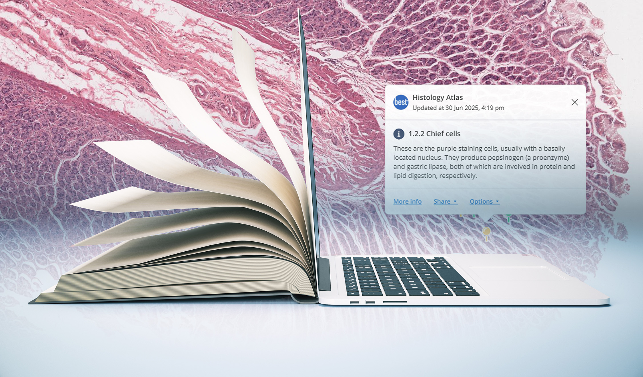
We’re excited to announce the release of the BEST Network Histology Atlas — a new, comprehensive online resource to support histology and histopathology teaching and learning.
Over the past twelve years, the BEST Network has reinvested subscription funding into building digital tools and resources for our global community of educators and students. From virtual slide hosting and image annotation to LTI integration and interactive “Drop a Pin” questions, these innovations have now come together in a long-requested format: an atlas.
The Histology Atlas brings together the best of what we’ve developed — now in one accessible, student-friendly package.
Slice currently hosts over 23,000 biomedical images, including 8,000+ virtual slides, used in histology and histopathology courses around the world. While this extensive image bank has served many institutions well, we heard consistent feedback from both academics and students:
The Histology Atlas answers both needs.
Designed as a self-paced, easy-to-navigate resource, the atlas includes:
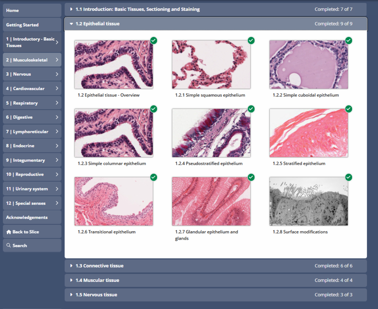
Detailed descriptions and diagrams
The atlas is organised by body system. Text based description are complemented by labelled diagrams and static images as well as embedded annotated virtual slides.

Annotated virtual slides
The annotated layers shared within the atlas support students with their examination of histological specimens compared to viewing images alone. Pins and/or regions mark multiple examples of features wherever possible to account for the normal variability within samples that will be seen by students while studying histology. Thanks to the integration of the virtual slides within the written content, features can be viewed in context at any magnification.

Integrated questions
“Drop a Pin” feature identification questions and MCQs are included on most pages to allow students to self-assess their understanding of a topic.
Whether used alongside a structured course or as a standalone study aid, the atlas provides flexible access for students at any stage of learning.
This project was made possible thanks to the contribution of material by The University of Queensland and in particular Dr Claire Aland who played a pivotal role in the development of this online Histology Atlas as part of her ongoing work with the BEST Network. We also acknowledge and thank Dr Kenneth Chan, who developed the original CD-based histology atlas in 1998 as part of his work at The University of Queensland. Special thanks to Dr Claire Aland, Professor Nicholas Hawkins, Dr Ildiko Erdelyi, Mr Benjamin Cai, Mr Max Kirsch, Dr Carolyn Cluderay and Dr Neill Sullivan for their contributions to and efforts in reviewing the Histology Atlas. We are incredibly grateful for the continued support of OpenLearning, whose cutting edge platform was the perfect place to integrate the content and the virtual slides on Slice. For a full list of our acknowledgements see within the atlas.
To access the Histology Atlas, login at www.best.edu.au and locate the Histology Atlas in the Account area or on the Slice navigation bar. Or watch our 1 minute explainer video.

If you have any questions about the Histology Atlas you're welcome to get in touch with me at s.dowdell@best.edu.au.
