Products


For generations of veterinary students at The University of Queensland, the anatomy "pot room" has been an important learning space. Shelves of preserved anatomical specimens, accompanied by printed reference cards containing labelled black-and-white photographs, provided students with the opportunity to study real anatomy up close.
Now, thanks to funding through The University of Queensland Faculty of Science Teaching and Learning Grant Scheme 2026, that resource is being transformed into a digital learning experience that students can access anywhere, anytime.
Preserving a Valuable Teaching Resource for the Future
One of the key drivers behind the project was the absence of a digital equivalent of the physical collection. While students could access the specimens in person at UQ's Gatton campus, the collection was unavailable outside of the Veterinary Anatomy pot room.
The project set out to create a comprehensive digital catalogue of the school's anatomical specimen collection.
Breathing New Life into an Ageing Collection
The project builds on a collection that has been used in veterinary teaching for many years. While the specimens themselves remain highly valuable educational tools, the supporting catalogue reflected an earlier era of educational technology.
The original reference materials consisted of printed black-and-white photographs with structures identified using letters and corresponding definitions on the reverse side of the card. Filing cabinets organised by body system housed hundreds of these cards.
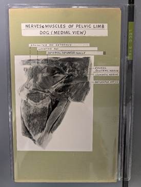
Example card showing labelled photograph
Capturing Anatomy in High Resolution
Creating digital versions of preserved specimens proved far more complex than simply taking photographs. With support from Dr Khama Kelman and her team at Murdoch University, who had established a workflow for high-depth of field specimen photography, the UQ project team set up professional photography equipment. This included specialised macro lenses, polarised lighting, light tents, image stacking software and colour calibration tools to produce detailed images with exceptional clarity and depth of field.
Many specimens required careful preparation before photography. Some pots needed cleaning or restoration, while others required replacement or top up of preservation liquid to improve visibility. However, working with preserved specimens introduced unique challenges as some containers had yellowed with age, while others contained scratches, dents or distortions that affected image quality. In some cases, specimens had shifted within the container over time, requiring careful manipulation to achieve the best possible photographic angles.
Veterinary student and project officer, Joshua Paech described one specimen as feeling like a puzzle game, carefully adjusting the position of structures within the fluid to recreate the intended anatomical view before it was photographed.
Solving Problems Through Innovation
One of the key technical challenges for the project involved presenting multiple views of a specimen in a way that remained intuitive for students. Anatomical structures are inherently three-dimensional, yet photographs capture only a single perspective.
As Paech discovered, some specimens required multiple cards to show different views and angles, making it difficult for students to build a complete understanding of the anatomy.
To overcome this, while photographing and preparing the digital images, Paech devised a Photoshop workflow that allowed multiple perspectives of the same specimen to be displayed on one image. This enables students to view and compare front, side and rear views without navigating between multiple images or files.
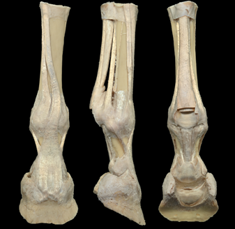
Fetlock joint, Horse showing multiple perspectives of the same specimen.
While staff acknowledge that digital images cannot fully replicate the experience of physically handling and rotating a specimen, they provide a significant step towards improving accessibility and understanding.
Learning Beyond the Pot Room
The digital specimens are being delivered through Slice, the resources are then integrated directly into Blackboard and other learning platforms. It also addresses a broader challenge within veterinary education. While veterinary schools across Australia and New Zealand recognise the value of sharing teaching resources and expertise, practical barriers often make this difficult. By creating and sharing this high-quality digital resource through Slice, UQ is hoping to support broader collaboration across institutions in the future.
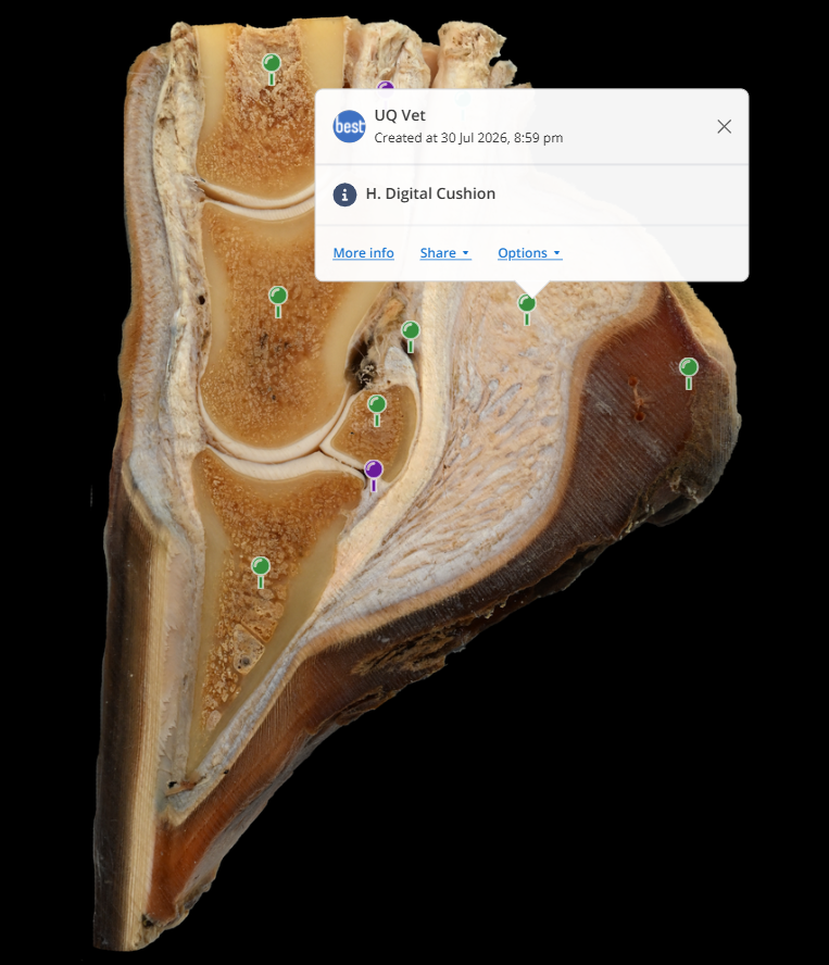
Using the Slice annotation tool, key muscles, vessels, nerves and other anatomical structures have been labelled within the high-resolution images. Students can zoom in on specimens, explore detailed annotations and switch layers on or off to support targeted revision.
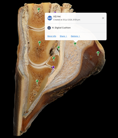
Horse Foot annotated to show bones, ligaments and tendons
An additional benefit has been the ability to update anatomical terminology. Some language used on the original reference cards no longer reflects current teaching practice, and staff can now update annotations quickly and consistently.
The resources have already been shared with students to support multiple anatomy subjects, with additional content continuing to be added. Although formal evaluation will begin following ethics approval and student surveys, early feedback has been overwhelmingly positive.
Paech recalls feeling particularly proud seeing students using the resources after they were released.
A Collection with Character
Among the collections available through Slice, this resource stands out for its consistently high-quality photography and exceptional depth of field. Of the hundreds of specimens photographed, a few stand out.
One favourite is affectionately known as "Meow Meow", a cat musculature specimen familiar to generations of veterinary students. For many UQ graduates, it is an instantly recognisable part of their anatomy studies.
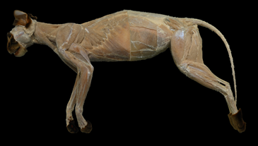
Deep muscles, Cat
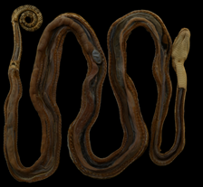
Entire body, Snake
Other highlights include reproductive anatomy specimens and comparative anatomy examples such as preserved snakes, demonstrating the diversity of the collection.
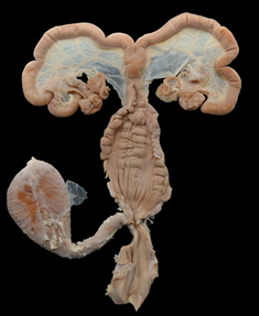
Pig female reproductive system
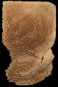
Reticulum, Zebu
The team's high-resolution imaging techniques have also captured levels of detail that would have been difficult to appreciate through older photographic methods, particularly in highly textured specimens such as the reticulum, a stomach compartment unique to ruminants.
Supporting Animal Ethics
The project also supports the universities commitment to the principles of the 3Rs: Replacement, Reduction and Refinement in animal use. By increasing the educational value and accessibility of existing specimens, the school can continue to maximise the learning benefit obtained from current resources while reducing the need for additional animal tissues in teaching.
What's Next?
While the current project has focused on creating a digital anatomy pot room, the team is already looking ahead with future plans to restore and digitise additional specimens that are currently not made available to students and the exploration of rotatable 3D anatomical models created through photogrammetry.
What began as a collection of preserved specimens and ageing filing cabinet cards is now a modern, accessible and collaborative digital learning environment, ensuring future generations of veterinary students can continue learning from these valuable resources wherever they are and setting up the opportunity for greater sharing of expertise and resources between veterinary schools.
Login to view the Digital Uplift project images. This project was supported by funding from The University of Queensland Faculty of Science - Teaching and Learning Grant Scheme 2026. Thank you to Dr Steph Bond and Joshua Paech for sharing their work and to the rest of the project team, Professor Paul Mills, Dr Claire Aland and Emma Greer.
The UQ project team would like to thank Dr Khama Kelman and the team from Murdoch University for sharing their techniques and advice.
